Our Company
Lazrostics is a team of Biomedical Engineering (BME) students dedicated to tackling post-surgical complications associated with
the treatment of esophageal cancer. Under the mentorship of Dr. Bernard Choi, Dr. William Tang, and Dr. Marc Fawaz, this project aims at developing the next generation endoscope that is capable of providing the end user with a
standard white-light view and a novel blood-flow sensing view. Sufficiently detailed real-time images of blood flow patterns beneath the surface of the
target under examination could provide valuable information about the health or disease states of the tissues. This will address the need to monitor
tissue blood perfusion in the esophagus after an esophagectomy (described in next section). Our device will provide clinicians a new way to monitor
and detect ischemic anastomosis.
The Problem
Esophagectomy
In 2012, there were about 456,000 new diagnoses of esophageal cancer worldwide with predicted deaths being 400,000 patients and the cancer carries a 5-year survival rate of 20 percent.
Esophagectomy is a complex surgical procedure that removes the cancerous sections of the esophagus, where the two ends are then sutured back together with
the bottom portion overlapping the top called an anastomosis.
 Surgical Procedure (Source: Mayo Clinic)
Diagnostic Complications
Post-surgical complications includes leaks, which occur when the two sutured ends give way, causing leakage which can lead to reduced blood flow and
septic shock. Esophogeal leakage rates can occur at 3-25% of all esophagectomies and carry a mortality rate of 30-60%.
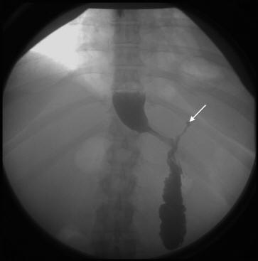 Anastomotic Leak (Source: Vizhul, Andrey et al. October 2011)
Clinical Unmet Need
Current detection methods for anastomotic leaks include CT scans and traditional endoscopes. Downsides of CT scan involves heavy radiation and injection
of harmful dyes that are associated with long-term negative effects, while traditional endoscopes can only evaluate tissue appearance but not blood flow.
Due to the high mortality rate associated with post-surgical complications in the treatment of esophageal cancer and the limited capabilities of
current detection techniques, it becomes critical to develop a device capable of sensing real-time blood flow in a minimally invasive manner.
Product
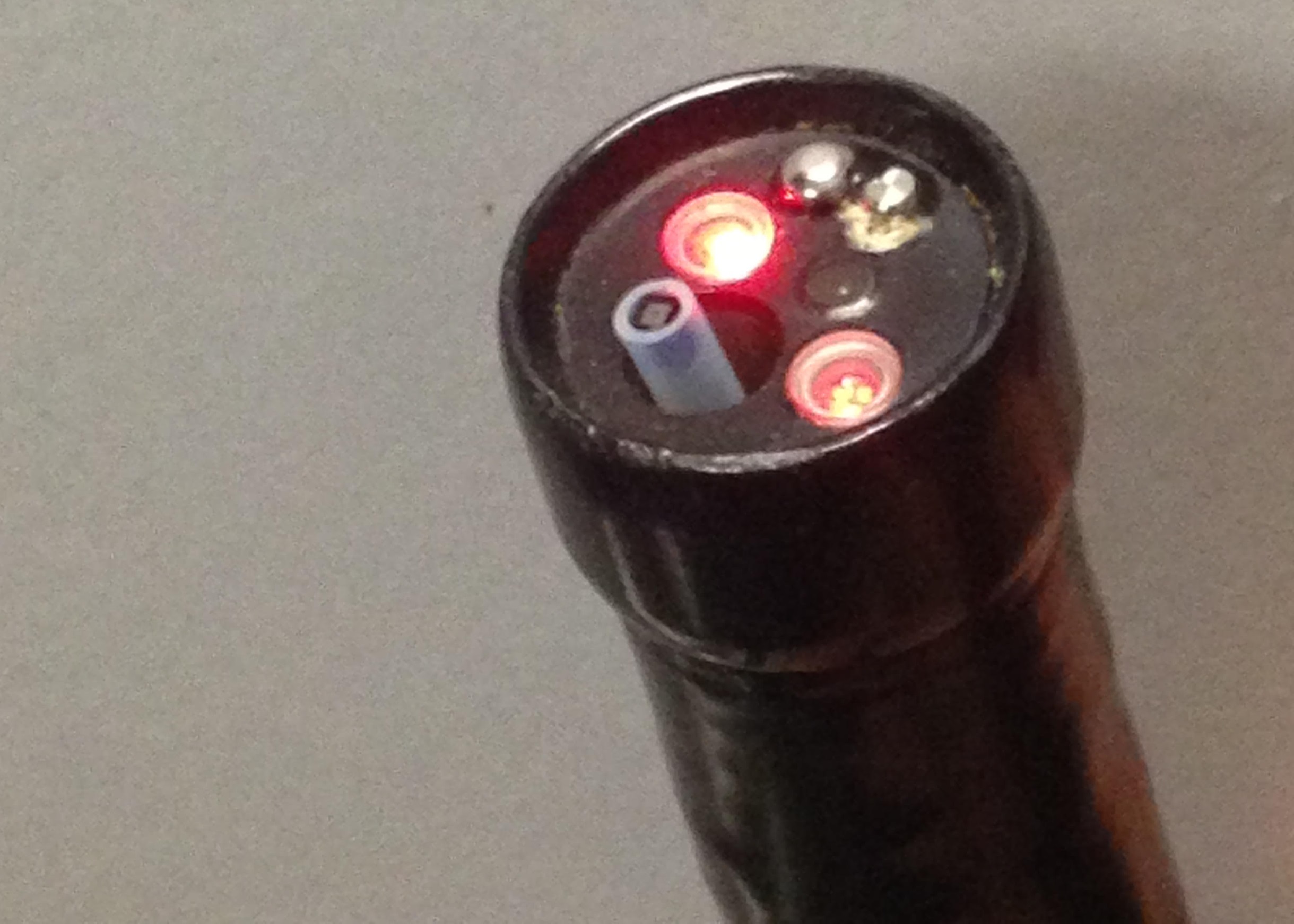 A typical endoscope holding the NanEye camera and using a red laser source
Concept Design
A typical endoscope shines a white light that reflects off the target and to be captured by the camera which sends the image to a viewing monitor.
In contrast, the ELSI uses a red laser to illuminate the target. The interference pattern of the laser, created when it interacts with the tissue,
is captured with the camera located in the endoscope tip. The captured data is then processed using MATLAB to present blood flow information on a viewing
monitor
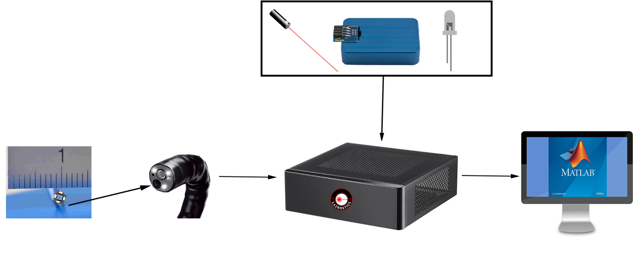 ELSI Flow Chart
The ELSI
Our device is called the Endoscopic Laser Speckle Imager(ELSI), which is made up of several
components such as a tiny camera that is placed in an endoscope tip, a red laser, a white LED, a data acquisition board, and a computer to process data.
The laser, LED, and data acquisition board are placed into a housing case which is machined to our specifications. Our device will have the option to
toggle between laser source and white light allowing us to acquire laser speckle and raw imaging respectively.
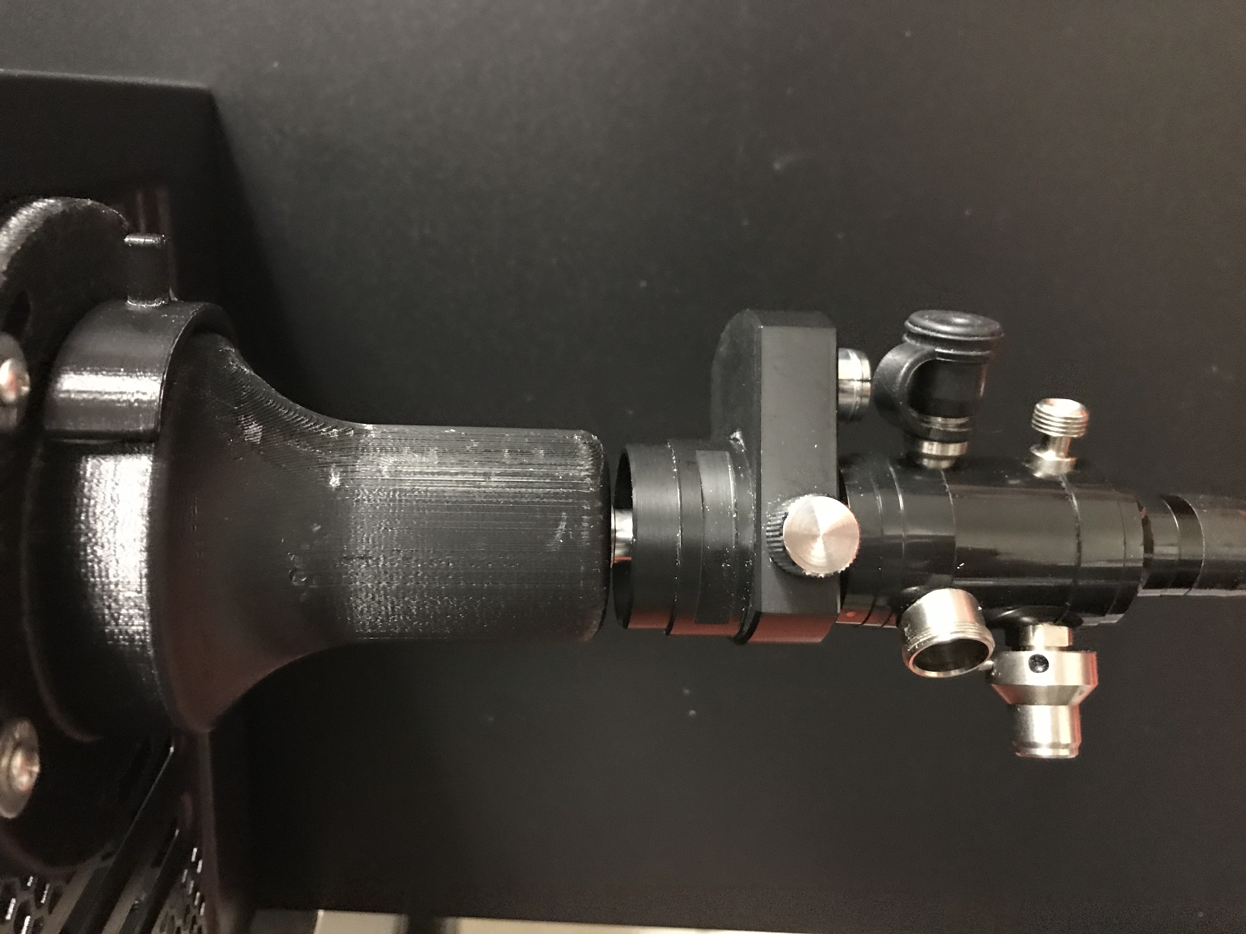
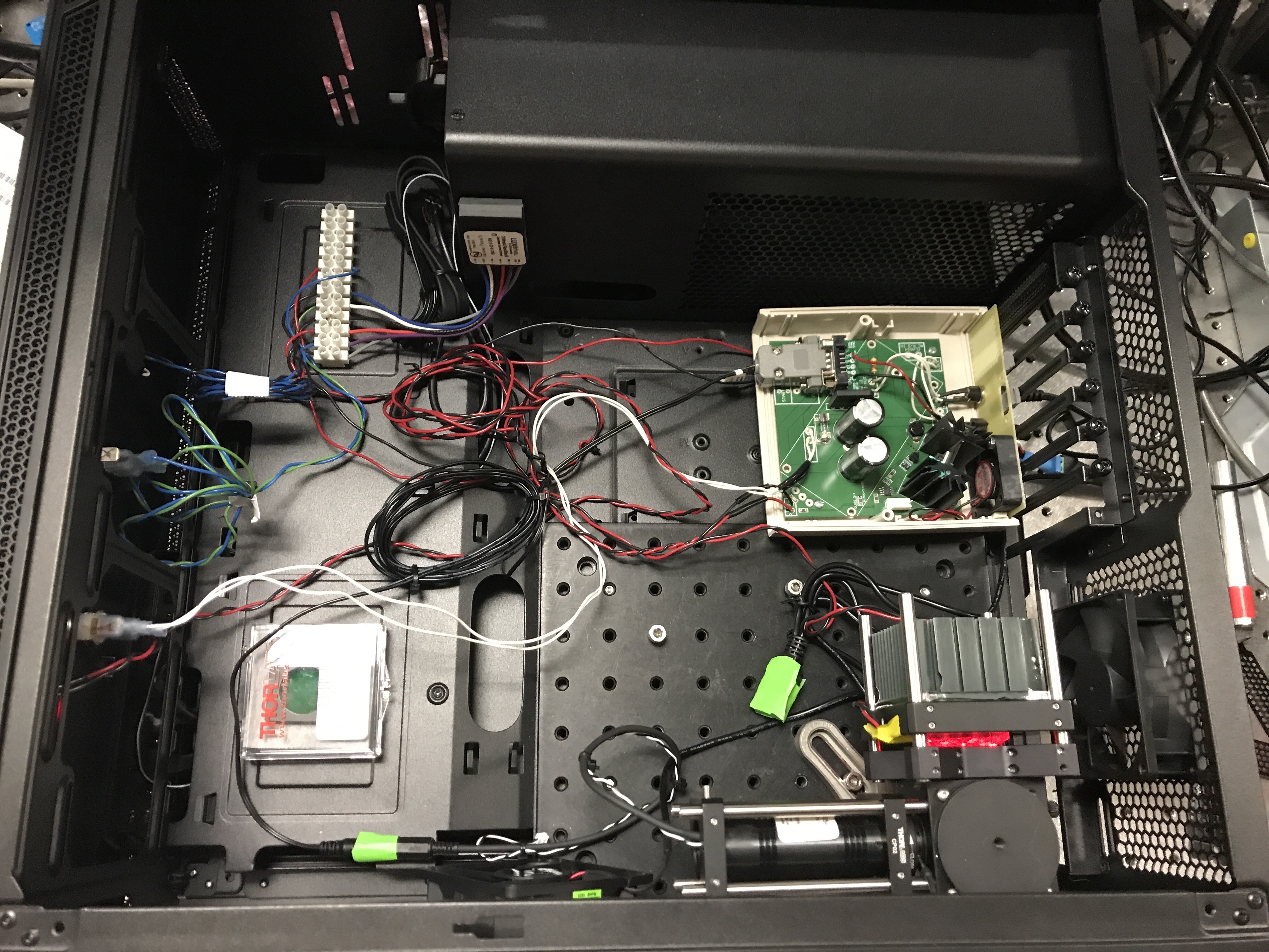 ELSI Box
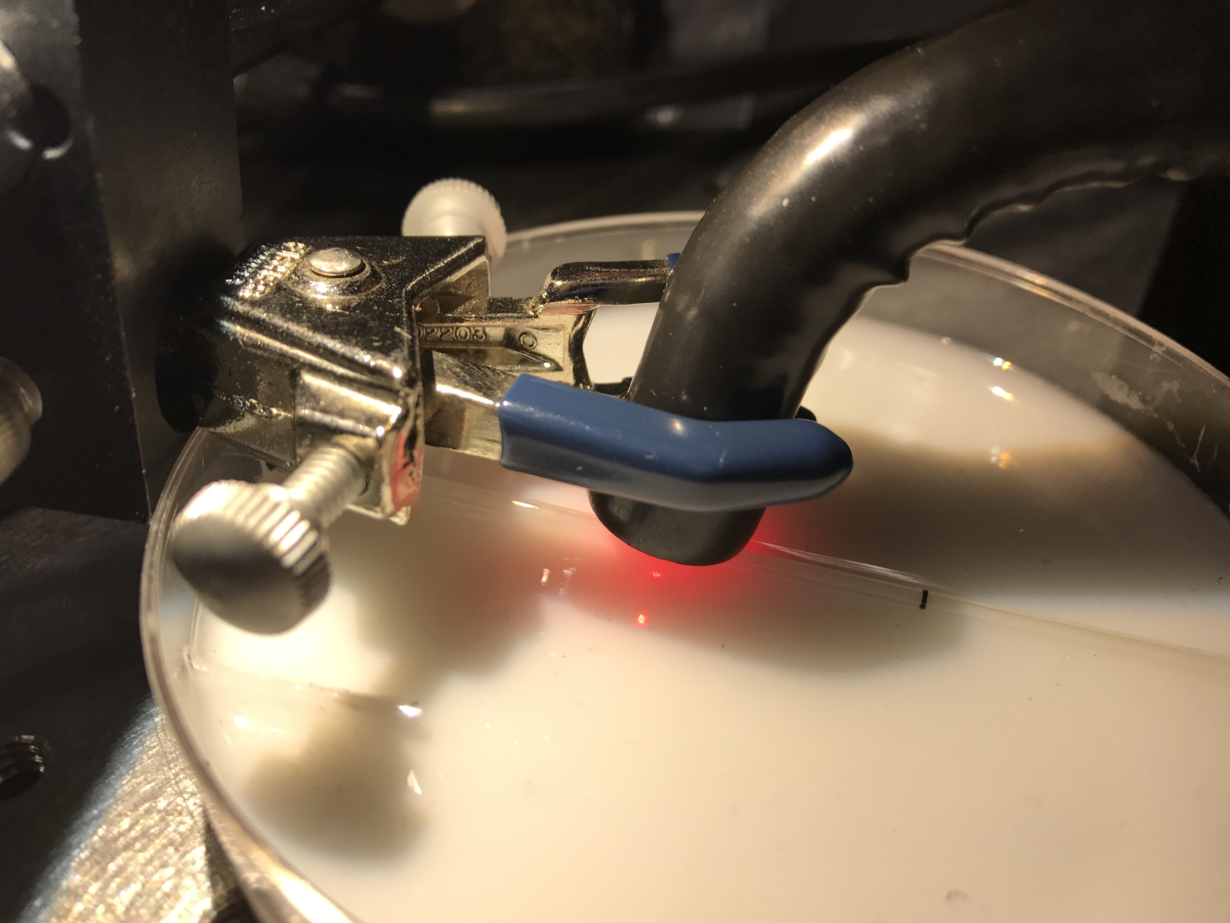
 Light Source Mount
 Indirect Competition Comparison
Device Testing
During development of a working prototype, our product needed to go through a series of flow phantom experiments and restricted blood flow tests.
The flow phantom test is made up of different components that have similar optical properties to the skin and of red blood cells to simulate blood flow in vitro.
When fluid is pumped through the phantom, our device should be able to detect the different flow speeds up to 5 mm/sec. If the speckle flow map is seen as too dark,
the problem would be with the red laser illumination or environmental setup. For the restricted blood flow test, a finger on a test subject’s hand will have a rubber
band to restrict blood flow where the colormap for the finger should appear blue. Finally, it will become necessary to test our product on porcine models to understand
the efficiency in vivo.
As a control, a CCD camera and external laser source are used together to visual blood flow on the flow phantom. This control experiment allows confirmation of the
efficiency of the matlab software and provides a baseline for our miniature camera/red laser experiment. Our device’s laser speckle image map must be similar to ones
that are shown using the CCD camera for further development and modifications of the device.
Our prototype demonstrates that adding blood flow sensing capabilities via a miniature camera has to potential to be a viable alternative to traditional methods of
blood flow sensing. Through in vivo testing the prototype was able to distinguish between various flow rate accurately and in real time.These features demonstrate the
device’s potential for numerous medical procedures and is not limited to post-surgical ischemic anastomosis. To maximize our ability to be competitive in this market,
our prototype needs to become more condensed and robust.
 Flow Phantom Experiment
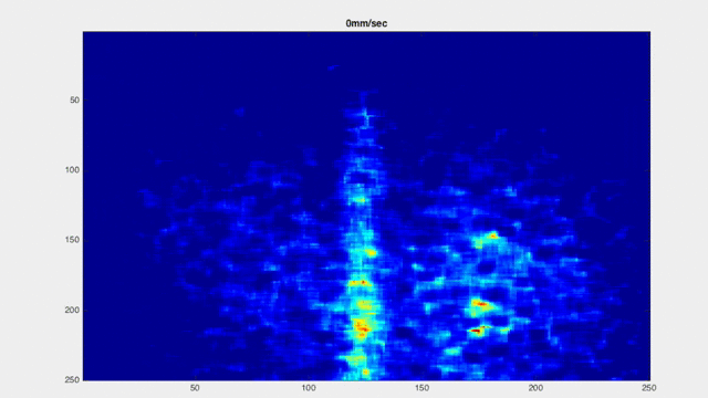 Speckle flow map at different speeds
Market Analysis
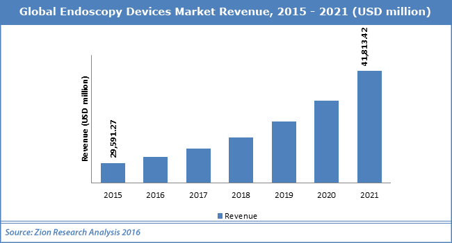 Source: Zion Market Research
Customers
Since our patient populations are those who undergo esophagectomies (esophogeal surgery), our customers are facilities
providing post-surgical care, such as hospitals, clinics, and primary care offices. Our device is designed to be used in any endoscopy procedure. Rising investments, funds, and grants by governments and hospitals worldwide for endoscopy equipment
has increased the endoscope market size. In 2016, North America is placed as the largest share of the endoscopy equipment market; however,
the Asia-Pacific market is projected to grow at the highest compound annual growth rate (CAGR) and will be a major revenue pocket for companies offering
endoscopy equipment.
Projected Plan
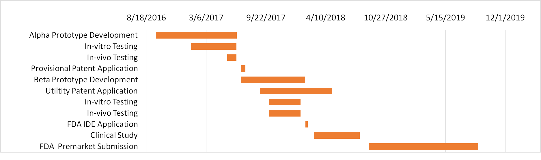 Commercialization Timeline
The endoscope market is predicted to exceed $30 billion by 2020.
After we have developed a working prototype, we will apply for the NIH Small Grant Program (R03) of up to $50,000/year to fund our verification and validation
research. Since the EG SCAN II and PeriCam PSI are our predicate devices, duration of the FDA approval process will be much shorter. After gaining FDA approval, we will
start a pilot production of 100 units, which we project to cost about $3,000 per device, which we will sell for $10,000. Considering that every diagnostic endoscopy procedure is reimbursed by Medicare at around $500 in-office and $1000 in an outpatient
hospital, our device is cost-effective to our customers.
Supporters


Lazrostics Team

Alexander Chang
Chief Executive Officer
Alexander has worked in the School of Medicine and has also worked under Dr. Andrei Shkel focusing on the vestibular endoscope.
Alexander is in charge of overseeing team work & external affairs.

Elijah De Guzman
Chief Operating Officer
Elijah is in charge of the internal works of the team, tracking project operations and organizing documents for our device.
Elijah is also in charge of designing computer models and currently works at a medical device startup.

Jonathan Abebe
Lead Software Engineer
Jonathan is experienced in Python, developing a GPS and game of Connect 4 against a server.
Jonathan is also a biological researcher and has presented his findings in conferences nationwide.
Miguel Bautista
Business Solutions Analyst
Miguel is in charge of the design aspects of the team, such as creating logos, videos, and writing reports.
Miguel currently works under Professor Michael D. Lee to analyze and plot NBA data.

Thien An Phuoc Ma
Chief Technology Officer
An Ma has experience developing the hardware & software for a low-cost ($250) handheld LSI system.
As Chief Technology Officer, An ensures the smooth hardware-software integration of our endoscopic LSI system.
Mentors

Bernard Choi, Ph.D.
Research Advisor
Dr. Choi’s research interests include the development and application of in vivo optical imaging methods
and technologies for monitoring of biological tissues in normal and diseased states, and for novel therapy discovery. He also leads
research efforts on the use of chemical agents to reduce the optical scattering of biological tissue. Dr. Choi provides mentorship towards
the research and design of our project.
Contact Us
Elements
i = 0;

while (!deck.isInOrder()) {
    print 'Iteration ' + i;
    deck.shuffle();
    i++;
}

print 'It took ' + i + ' iterations to sort the deck.';